Acid-Fast Staining (Ziehl-Neelsen and Kinyoun) Procedures

Ziehl-Neelsen (Hot Stain) Procedure
  1. Prepare a smear consisting of a mixture of E. coli and M. smegmatis.
  2. Allow the smear to air dry and then heat-fix (see figure 7.1).
  3. Place the slide on a hot plate that is within a chemical hood (with the exhaust fan on), and cover the smear with a piece of paper towelingthat has been cut to the same size as the microscope slide. Saturate the paper with Ziehl’s carbolfuchsin (figure a). Heat for 3 to 5 minutes. Do not allow the slide to dry out, and avoid excess flooding! Also, prevent boiling by adjusting the hot plate to a proper temperature. A boiling water bath with a staining rack or loop held 1 to 2 inches above the water surface also works well. (Instead of using a hot plate to heatdrive the carbolfuchsin into the bacteria, an alternate procedure is to cover the heat-fixed slide with a piece of paper towel. Soak the towel with the carbolfuchsin and heat, well above a Bunsen burner flame.)
  4. Remove the slide, let it cool, and rinse with water for 30 seconds (figure b).
  5. Decolorize by adding acid-alcohol drop by drop until the slide remains only slightly pink. Thisrequires 10 to 30 seconds and must be done carefully (figure c).
  6. Rinse with water for 5 seconds (figure d).
  7. Counterstain with alkaline methylene blue for about 2 minutes (figure e).
  8. Rinse with water for 30 seconds (figure f).
  9. Blot dry with bibulous paper (figure g).
  10. There is no need to place a coverslip on the stained smear. Acid-fast organisms stain red; the background and other organisms stain blue or brown.


Kinyoun (Cold Stain) Procedure
(This may be used instead of or in addition to the Ziehl-Neelsen procedure.)
  1. Heat-fix the slide as previously directed.
  2. Flood the slide for 5 minutes with carbolfuchsin prepared with Tergitol No. 7 (heat is not necessary).
  3. Decolorize with acid-alcohol and wash with tap water. Repeat this step until no more color runs off the slide.
  4. Counterstain with alkaline methylene blue for 2 minutes. Wash and blot dry.
  5. Examine under oil. Acid-fast organisms stain red; the background and other organisms stain blue.

Gram Stain

  1. Prepare heat-fixed smears of E. coli, S. aureus, and the mixture of E. coli and S. aureus (see figure 7.1).
  2. Place the slides on the staining rack.
  3. Flood the smears with crystal violet and let stand for 30 seconds (figure a).
  4. Rinse with water for 5 seconds (figure b).
  5. Cover with Gram’s iodine mordant and let stand for 1 minute (figure c).
  6. Rinse with water for 5 seconds (figure d).
  7. Decolorize with 95% ethanol for 15 to 30 seconds. Do not decolorize too long. Add the decolorizer drop by drop until the crystal violet fails to wash from the slide (figure e). Alternatively, the smears may be decolorized for 30 to 60 seconds with a mixture of isopropanol-acetone (3:1 v/v).
  8. Rinse with water for 5 seconds (figure f ).
  9. Counterstain with safranin for about 60 to 80 seconds (figure g). Safranin preparations vary considerably in strength, and different staining
  10. times may be required for each batch of stain. (If you are color-blind, use Bismark brown stain rather than safranin.)
  11. Rinse with water for 5 seconds (figure h).
  12. Blot dry with bibulous paper (figure i) and examine under oil immersion. Gram-positive organisms stain blue to purple; gram-negative organisms stain pink to red. There is no need to place a coverslip on the stained smear. See figure bellow for an example of gram-positive and gram negative bacteria.

Smear Preparation and Simple Staining

Bacterial smear and staining preparation
Smear procedure:
  1. With the pen (permanent ink)/pencil, mark the name of the bacterial culture in the far left corner on each slides.
  2. For the broth culture, shake the culture tube and, with an inoculating loop, aseptically (see figure bellow) transfer 1 to 2 loopfuls of bacteria to the center of the slide. Spread this out to about a 1/2-inch area. When preparing a smear from a slant or plate, place a loopful of water in the center of the slide. With the inoculating needle, aseptically pick up a very small amount of culture and mix into the drop of water. Spread this out as above.
  3. Allow the slide to air dry, or place it on a slide warmer (see figure bellow).
  4. Pass the slide through a Bunsen burner flame three times to heat-fix and kill the bacteria.


Simple Staining:
  1. Place the fixed smears on a staining loop or rack over a sink or other suitable receptacle
  2. Stain one slide with alkaline methylene blue for 1 to 1,5 minutes, or carbolfuchsin for 5 to 10 seconds, or with crystal violet for 20 to 30 seconds.
  3. Wash stain off slide with water for a few seconds (see figure bellow).
  4. Blot slide dry with bibulous paper.Be careful not to rub the smear when drying the slide because this will remove the stained bacteria.
  5. Examine under the oil immersion lens.

Preparation of Streak Plate

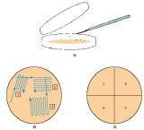
Arrows indicate motion of the loop. In b, flame and cool the loop between 1 and 2, 2 and 3, and 3 and the end of the streak. The goal is to thin the numbers of bacteria growing in each successive area of the plate as it is rotated and streaked so that well isolated colonies will appear in quadrant 3.